The horse’s hoof is a miracle of engineering. It contains a whole host of structures which, when healthy, operate in equilibrium with each other to form a hoof capsule which is able to withstand huge forces, utilising energy to assist with forward movement while providing protection to the sensitive structures beneath.
This is not a definitive guide, but it will allow the horse owner to understand more about this incredible structure.
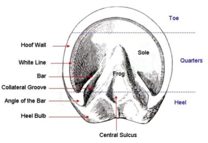
EXTERNAL STRUCTURES
This newly trimmed section of hoof gives you a good idea of what the different structures of the hoof wall and sole look like. 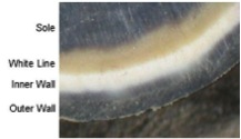 On a white foot, the differences are much less easy to spot.
Sole
The sole is the area inside the white line, but not including the bars and frog. It’s primary function is to protect the sensitive structures beneath the sole. However, the outer perimeter of sole around the toe also provides support, sharing some of the weight of the horse with the hoof wall.
White Line
Commonly referred to as the white line, although this is very misleading, not only because it is actually yellowish but also because it is next to the white inner wall of the hoof. This often causes people to misinterpret the white line as inner wall, so it is sometimes called the Golden Line – more accurate description that was commonly used in the 1800s. The purpose of the Golden Line is to join the sole to the inner wall of the hoof and to seal off the border of the pedal bone to protect it from bacterial infiltration. It creates a shallow crease at the bottom of the hoof which fills with dirt, aiding with traction.
Inner Wall
The inner hoof wall is usually white (unlike the outer wall, it does not contain pigment). It is more pliable than the outer wall due to it having a higher moisture content which enables the inner wall to stretch more as the outer wall moves, ensuring the inner workings of the hoof are protected from too much shock as well as allowing the pedal bone and the outer wall to move in different ways without losing strength of attachment.
Outer Wall
The outer hoof wall is pigmented and is much stronger than the inner wall. Its purpose is to bear the weight of the horse, protect the internal structures from harm and to act like a spring, storing and releasing energy during the different phases of the stride to help propel the horse along. A healthy outer wall will be slightly thicker at the toe and have no growth rings or cracks. It will be almost impermeable, meaning water or any other things which come in contact with it will not be able to penetrate the wall. If, however, an outer wall is damaged – either through injury or nutritional imbalance, substances which come into contact with it will seep through.
Bar
The bar is an extension of the hoof wall which runs along the side of the frog, terminating approximately half way along the frog. Its primary purpose is to control the movement of the back of the hoof, adding strength to the heel area and protecting it from excess distortion. It should have a high ratio of pliable inner wall to ensure it can move correctly as the heel moves. More recent research has found that the bar also produces some of the material that makes up the sole.
Angle of the Bar
Commonly known as the heel, although this description can be misleading. This area is designed to receive the initial impact of the horse’s stride and a healthy angle of the bar comprises mainly of pliable inner wall, enabling it to dissipate excess shock with ease. This area plays a major role in supporting the weight of the horse and it is important that it remains correctly balanced.
Collateral Groove
This is the groove that runs along either side of the frog. The outer wall of the groove is made up of the wall of the bar and sole and the wall on the other side comprises the wall of the frog.
Frog
One of the most important, but often neglected structures of the horse’s hoof. It should be wide and substantial and made up of thick, leathery material. An unhealthy frog is vulnerable to infection which, if left untreated, can lead to significant loss of structure in the back of the hoof causing severe lameness.
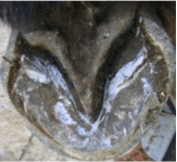 The frog works in concert with the coronet band, the bars and the sole to provide resistance to distortion of the hoof capsule during the stride. Pressure placed upon the frog directly influences the health of the digital cushion above it. The frog stay (triangular piece cut out of the sole that the frog sits in) allows independent movement at the heels as the horse lands on uneven ground. The frog also plays a part in protecting the sensitive structures beneath, providing traction, assisting circulation and absorbing shock. It also contains many nerves which enable the horse to feel what it is standing on and be aware of where its feet are in relation to the rest of its body (proprioception).
In the centre of the frog, towards the back of the foot is the central sulcus. A healthy sulcus is wide and shallow, but if the frog is weak and narrow it can become a deep crease which is a haven for bacteria and fungus.
Coronet Band
In the UK the coronet band is thought to be so called because coronet means “crown”. In other parts of the world the phrase “Coronary band” is often used – coronary meaning “pertaining to the heart”. This is a very tough, vascular structure which sits at the top of the hoof wall. It has two very important functions. Firstly it produces the tubules of the outer hoof wall. Secondly, it is incredibly strong and acts as a band of support to add strength to the internal structures as the hoof distorts during the stride.
Periople
This is a protective covering for the area of newly formed hoof wall just below the coronet band. In the early stages, this horn material is quite soft – deliberately so because it helps to prevent the coronet band becoming bruised as shock is transferred upwards through the hoof wall during the weight bearing phase of the stride. The periople covers this horn to provide protection.
INTERNAL STRUCTURES
There are two and a bit bones inside the hoof. The Pedal bone, the Navicular bone and the bottom part of the Short Pastern bone.
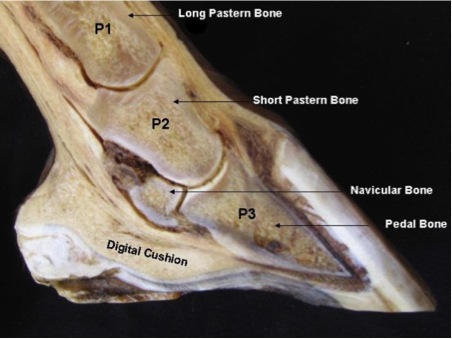
Pedal Bone
The large bone inside the hoof capsule is known as the Pedal bone or Coffin bone. Its shape provides a framework for the shape of the hoof capsule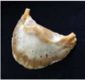 itself.
The Pedal bone provides strength and stability to the hoof and acts as a framework to hold other structures in place. It does not have a medulla (bone marrow) and has an unusually high density of tiny blood vessels running through it. Surrounding the wall of the bone is the laminae which hold the wall to the bone and produce some of the intertubular horn of the hoof wall. Underneath, the bone is covered in solar corium which produces the sole. At the back, the bone attaches to cartilage which forms a large portion of the back of the hoof . Tendons and ligaments are attached to this bone and a dense network of blood vessels run around and through it.
Navicular Bone
Navicular bone
This is another bone which is hard to visualise when viewed in cross section. It is thought to have derived its name because it is shaped like a boat.It is also known as the Distal Sesamoid bone (distal meaning furthermost from the body, sesamoid meaning embedded within a tendon). The navicular bone is not actually embedded in a tendon, but it does sit just inside the back of the pedal bone and the deep digital flexor tendon passes over it. It prevents over-articulation of the joint of the pedal bone, maintains a constant angle of insertion of the Deep Flexor Tendon into the back of the Pedal bone and allows for additional tilt within the coffin joint when navigating uneven surfaces.
Short Pastern Bone
Also known as the Middle Phalanx, the short pastern bone sits on top of the articulating joint of the pedal bone and underneath the long pastern bone. Only the bottom portion of this bone extends as far as the hoof capsule.
Digital Cushion
The digital cushion sits just behind the pedal bone and above the sensitive frog. It plays a vital role in the absorption of shock. In an improperly functioning foot, the digital cushion atrophies and becomes “fatty” as opposed to springy, cartilaginous material, inhibiting its ability to absorb shock. The shape and health of the digital cushion will influence the angle of the Pedal Bone. “Flat footed” horses (ie, those whose pedal bones lie flat instead of being tilted slightly on their nose) often have severely atrophied digital cushions.
Coriums
A corium is a vascular structure which manufactures hoof horn. For instance, the solar corium will produce the sole and the frog corium produces the frog. The coronet band contains a corium which produces the tubules and intertubular horn of the hoof wall, whereas more intertubular horn is manufactured in the corium surrounding the pedal bone (also known as the dermal layer or laminae). The perioplic corium sits under the coronet band and produces the periople.
Lateral/Ungual Cartilage
 The lateral cartilages are located both above and below the coronet band, extending around the front, the sides and back of the hoof. Below the coronet band they extend out over the digital cushion and attach to the back of the pedal bone. The horn producing corium of the inner hoof wall attaches to the lateral cartilages at the back of the hoof where the pedal bone does not reach. These cartilages provide resistance as the pedal bone descends during weight bearing, regulating the amount of pressure applied to the coriums. They also help to suspend the pedal bone in the correct position as well as acting as a spring, storing and releasing energy during locomotion.
Blood supply
The hoof is heavily supplied with blood through the two arteries which run down the back of the leg and into the foot. 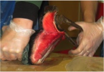 The pedal bone itself has an unusually high density of blood vessels within it. The photograph shows the laminae which keep the hoof wall tightly bonded to the internal structures. The blood pumping around the foot has many vital uses. It supplies nutrients to allow growth of new horn tissue, assists with damping impact shock and helps to regulate hoof temperature.
This is just a taster of what an amazing and complex structure the equine hoof is. If you would like to learn more about the anatomy of the equine hoof, and the way it functions I suggest you read the following:
“Practical Guide to Lameness in Horses”, Ted S Stashak, ISBN 0-683-07985-9
“The Equine Distal Limb”, Jean-Marie Denoix, ISBN 978-1840760019
“Equine Podiatry”. Andrea E Floyd, Richard A Mannesmann, ISBN: 978-0-7216-0383-4
“Anatomy of the Horse”, Klaus-Dieter Budras, W.O. Sack, Sabine Röck, ISBN 978-3-89993-044-3
This article has been written and kindly donated by Jayne Hunt (www.healthyhooves.co.uk). Jayne is a professional Equine Podiatrist and trainer who specialises in Equine Anatomy and Physiology, which she teaches to students studying the two year Diploma in Equine Podiatry course with Equine Podiatry Training Ltd www.eptrain.co.uk.